41 Year Old Male Came with Abdominal Pain, Bloating and Jaundice.
Findings:
MRI Liver
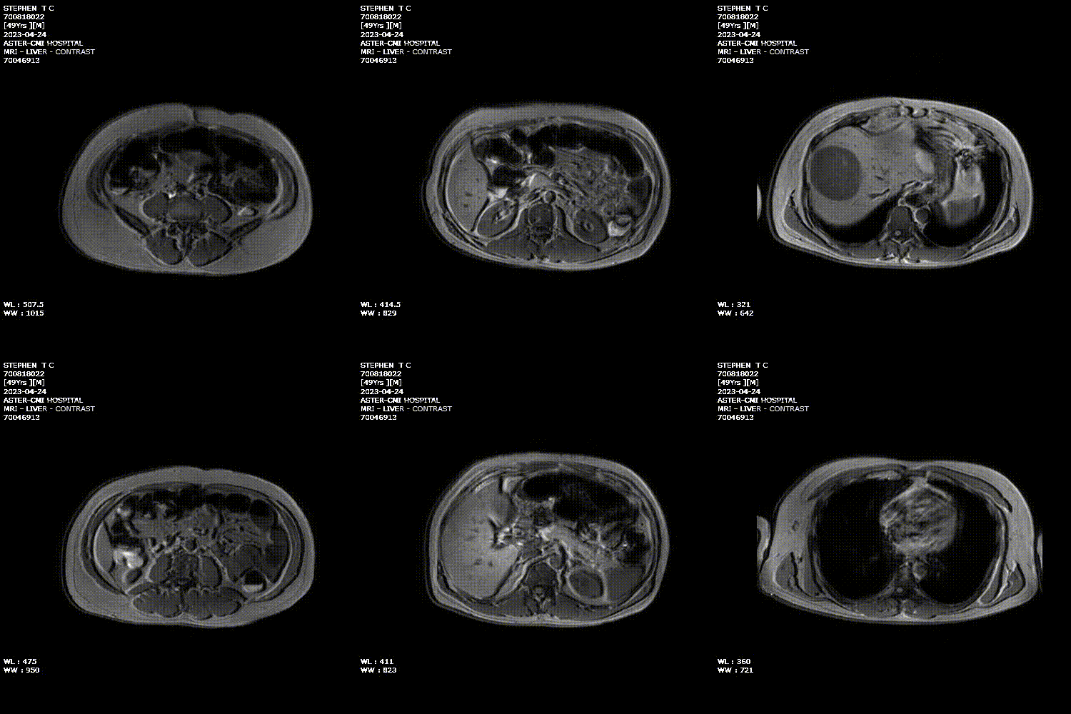


- Well-defined round to oval T2 hyperintense and T1 hypointense lesion in segment 8 of the liver which showed diffusion restriction. Multiple T2 hyperintense septae and a few tiny foci of T1 hyper intensities were noted within the lesion. No arterial enhancement was seen. Faint progressive enhancement in the venous and delayed phase of imaging was seen predominately in the periphery of the lesion with enhancement of the septae within the lesion.
- There was no evidence of intra hepatic biliary radicular dilatation.
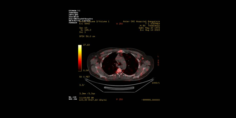
Follow-up with PETCT after 4 months showed:
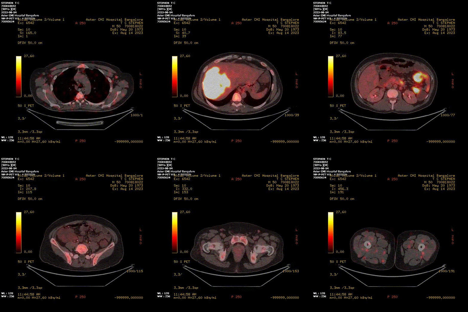
- Interval increase in the size of the liver lesion with FDG uptake. The lesion was seen extending along the right hepatic duct, medially extending to confluence, resulting in significant IHBRD on the left side. The lesion was seen extending into the right portal vein with evidence of thrombus.
- Another metabolically active hypodense lesion was noted in the right kidney.
- Metabolic active irregular wall thickening involving jejunum.
- Multiple metabolically active retroperitoneal lymph nodes were also noted.
- FDG uptake is also seen in soft tissue density deposit in subhepatic region.
DIAGNOSIS:
- Multisystem lymphoma
DISCUSSION:
Lymphomas can be restricted to the lymphatic system or can arise as extra nodal disease. This, along with variable aggressiveness results in diverse imaging appearance. Secondary hepatic involvement with lymphoma is much more common than primary hepatic lymphoma. The kidneys are the most common abdominal organs affected by lymphoma.
Differential Diagnosis:
- Hepatocellular carcinoma with metastasis- HCC will show arterial enhancement.
- Renal cell carcinoma-Usually heterogeneous with vascular invasion.
- Embryonal sarcoma of the liver- Metastases are rare and when seen, are most frequently found in the lungs, pleura, peritoneum, and bone.
Contributing Authors:
- Dr Saarah Khan - ASTER CMI Hospital, Bangalore
- Dr Shyam - ASTER CMI Hospital, Bangalore
- Dr Swathi - ASTER CMI Hospital, Bangalore
- Dr Sudhir Kale - ASTER CMI Hospital, Bangalore
REFERENCES:
- Bach AG, Behrmann C, Holzhausen HJ et al (2012) Prevalence and imaging of hepatic involvement in malignant lymphoproliferative disease.
- Buchpiguel CA (2011) Current status of PET/CT in the diagnosis and follow up of lymphomas. Rev Br Hematol Hemoter 33:140–147.

AMI Expertise - When You Need It, Where You Need It.
Partner With Us

